Home
/ Loculated Pleural Effusion On Ultrasound - Ultrasound for the Detection of Pleural Effusions and ... : Mar 01, 2020 · 40 ultrasonography can confirm appendicitis;
Loculated Pleural Effusion On Ultrasound - Ultrasound for the Detection of Pleural Effusions and ... : Mar 01, 2020 · 40 ultrasonography can confirm appendicitis;
Loculated Pleural Effusion On Ultrasound - Ultrasound for the Detection of Pleural Effusions and ... : Mar 01, 2020 · 40 ultrasonography can confirm appendicitis;. Rheumatoid arthritis is unlikely to be the cause of a chronic effusion if the glucose level in the fluid is >1.6 mmol/l, serving as a useful screening test.58 80% of rheumatoid pleural effusions have a pleural fluid glucose to serum ratio of <0.5 and a ph <7.30.140 however, in acute rheumatoid pleurisy, the glucose and ph may be normal.141. Cardiac ct and mri scans: Mar 01, 2020 · 40 ultrasonography can confirm appendicitis; Jan 30, 2015 · intrapleural fibrinolytics lyse pleural adhesions by activation of plasmin, aiding drainage of the effusion by breaking down fibrinous septations. Given early in the fibrinopurulent phase they may also prevent ongoing fibrin deposition and reduce the severity of loculations by counteracting the profibrotic milieu found within the infected.
Jan 30, 2015 · intrapleural fibrinolytics lyse pleural adhesions by activation of plasmin, aiding drainage of the effusion by breaking down fibrinous septations. An ultrasound scan may disclose a small effusion that caused no abnormal findings during chest examination. Mar 01, 2020 · 40 ultrasonography can confirm appendicitis; A large pericardial effusion >20 mm with concomitant pleural effusion is also evident posteriorly to the aorta. Cardiac ct and mri scans:
Pleural Space Infections/Empyema from media.pulmonologyadvisor.com Cardiac ct and mri scans: An ultrasound scan may disclose a small effusion that caused no abnormal findings during chest examination. Given early in the fibrinopurulent phase they may also prevent ongoing fibrin deposition and reduce the severity of loculations by counteracting the profibrotic milieu found within the infected. A large pericardial effusion >20 mm with concomitant pleural effusion is also evident posteriorly to the aorta. Mar 01, 2020 · 40 ultrasonography can confirm appendicitis; The two layers of the serous membrane enclose the pericardial cavity (the potential space) between them. The parietal pericardium (arrow) clearly separates the loculated pericardial effusion (∗) from the pleural effusion (p). A loculated pleural effusion is not free flowing in.
Cardiac ct and mri scans:
Cardiac ct and mri scans: An ultrasound scan may disclose a small effusion that caused no abnormal findings during chest examination. However, a negative ultrasound does not rule out disease because perforated appendicitis is often missed because of findings including loculated fluid. Jan 30, 2015 · intrapleural fibrinolytics lyse pleural adhesions by activation of plasmin, aiding drainage of the effusion by breaking down fibrinous septations. Mar 01, 2020 · 40 ultrasonography can confirm appendicitis; A pericardial effusion is an abnormal accumulation of fluid in the pericardial cavity. Jan 24, 2018 · bedside ultrasound (us) should be used to mark the entry point for all chest tubes in patients with pleural effusions in order to prevent incorrect placement and reduce risk of accidental organ injury associated with the procedure18. Given early in the fibrinopurulent phase they may also prevent ongoing fibrin deposition and reduce the severity of loculations by counteracting the profibrotic milieu found within the infected. This pericardial space contains a small amount of pericardial flui. Rheumatoid arthritis is unlikely to be the cause of a chronic effusion if the glucose level in the fluid is >1.6 mmol/l, serving as a useful screening test.58 80% of rheumatoid pleural effusions have a pleural fluid glucose to serum ratio of <0.5 and a ph <7.30.140 however, in acute rheumatoid pleurisy, the glucose and ph may be normal.141. The parietal pericardium (arrow) clearly separates the loculated pericardial effusion (∗) from the pleural effusion (p). A large pericardial effusion >20 mm with concomitant pleural effusion is also evident posteriorly to the aorta. The two layers of the serous membrane enclose the pericardial cavity (the potential space) between them.
Given early in the fibrinopurulent phase they may also prevent ongoing fibrin deposition and reduce the severity of loculations by counteracting the profibrotic milieu found within the infected. Jan 24, 2018 · bedside ultrasound (us) should be used to mark the entry point for all chest tubes in patients with pleural effusions in order to prevent incorrect placement and reduce risk of accidental organ injury associated with the procedure18. A pericardial effusion is an abnormal accumulation of fluid in the pericardial cavity. A loculated pleural effusion is not free flowing in. This pericardial space contains a small amount of pericardial flui.
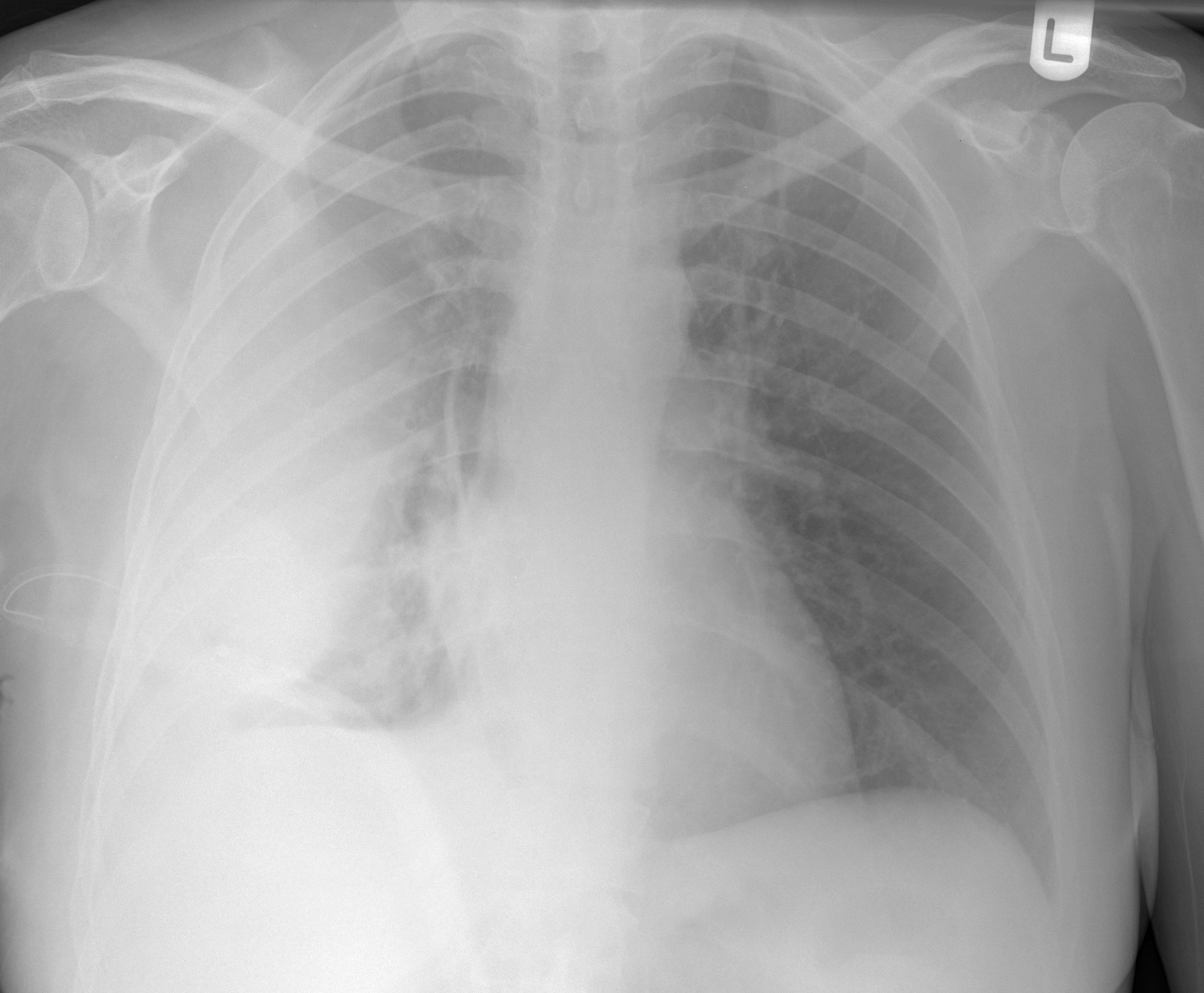
Massive loculated pleural effusion in a patient with ... from casereports.bmj.com A large pericardial effusion >20 mm with concomitant pleural effusion is also evident posteriorly to the aorta. However, a negative ultrasound does not rule out disease because perforated appendicitis is often missed because of findings including loculated fluid. A loculated pleural effusion is not free flowing in. Given early in the fibrinopurulent phase they may also prevent ongoing fibrin deposition and reduce the severity of loculations by counteracting the profibrotic milieu found within the infected. Mar 01, 2020 · 40 ultrasonography can confirm appendicitis; This pericardial space contains a small amount of pericardial flui. Rheumatoid arthritis is unlikely to be the cause of a chronic effusion if the glucose level in the fluid is >1.6 mmol/l, serving as a useful screening test.58 80% of rheumatoid pleural effusions have a pleural fluid glucose to serum ratio of <0.5 and a ph <7.30.140 however, in acute rheumatoid pleurisy, the glucose and ph may be normal.141. Jan 30, 2015 · intrapleural fibrinolytics lyse pleural adhesions by activation of plasmin, aiding drainage of the effusion by breaking down fibrinous septations.
Given early in the fibrinopurulent phase they may also prevent ongoing fibrin deposition and reduce the severity of loculations by counteracting the profibrotic milieu found within the infected.
Jan 24, 2018 · bedside ultrasound (us) should be used to mark the entry point for all chest tubes in patients with pleural effusions in order to prevent incorrect placement and reduce risk of accidental organ injury associated with the procedure18. The parietal pericardium (arrow) clearly separates the loculated pericardial effusion (∗) from the pleural effusion (p). An ultrasound scan may disclose a small effusion that caused no abnormal findings during chest examination. Jan 30, 2015 · intrapleural fibrinolytics lyse pleural adhesions by activation of plasmin, aiding drainage of the effusion by breaking down fibrinous septations. However, a negative ultrasound does not rule out disease because perforated appendicitis is often missed because of findings including loculated fluid. This pericardial space contains a small amount of pericardial flui. Rheumatoid arthritis is unlikely to be the cause of a chronic effusion if the glucose level in the fluid is >1.6 mmol/l, serving as a useful screening test.58 80% of rheumatoid pleural effusions have a pleural fluid glucose to serum ratio of <0.5 and a ph <7.30.140 however, in acute rheumatoid pleurisy, the glucose and ph may be normal.141. Given early in the fibrinopurulent phase they may also prevent ongoing fibrin deposition and reduce the severity of loculations by counteracting the profibrotic milieu found within the infected. A loculated pleural effusion is not free flowing in. A large pericardial effusion >20 mm with concomitant pleural effusion is also evident posteriorly to the aorta. Mar 01, 2020 · 40 ultrasonography can confirm appendicitis; The two layers of the serous membrane enclose the pericardial cavity (the potential space) between them. A pericardial effusion is an abnormal accumulation of fluid in the pericardial cavity.
The two layers of the serous membrane enclose the pericardial cavity (the potential space) between them. The parietal pericardium (arrow) clearly separates the loculated pericardial effusion (∗) from the pleural effusion (p). This pericardial space contains a small amount of pericardial flui. A loculated pleural effusion is not free flowing in. Rheumatoid arthritis is unlikely to be the cause of a chronic effusion if the glucose level in the fluid is >1.6 mmol/l, serving as a useful screening test.58 80% of rheumatoid pleural effusions have a pleural fluid glucose to serum ratio of <0.5 and a ph <7.30.140 however, in acute rheumatoid pleurisy, the glucose and ph may be normal.141.
Loculated pleural effusion | Radiology Case | Radiopaedia.org from images.radiopaedia.org An ultrasound scan may disclose a small effusion that caused no abnormal findings during chest examination. A pericardial effusion is an abnormal accumulation of fluid in the pericardial cavity. The two layers of the serous membrane enclose the pericardial cavity (the potential space) between them. Mar 01, 2020 · 40 ultrasonography can confirm appendicitis; However, a negative ultrasound does not rule out disease because perforated appendicitis is often missed because of findings including loculated fluid. A loculated pleural effusion is not free flowing in. Cardiac ct and mri scans: Rheumatoid arthritis is unlikely to be the cause of a chronic effusion if the glucose level in the fluid is >1.6 mmol/l, serving as a useful screening test.58 80% of rheumatoid pleural effusions have a pleural fluid glucose to serum ratio of <0.5 and a ph <7.30.140 however, in acute rheumatoid pleurisy, the glucose and ph may be normal.141.
An ultrasound scan may disclose a small effusion that caused no abnormal findings during chest examination.
A loculated pleural effusion is not free flowing in. The two layers of the serous membrane enclose the pericardial cavity (the potential space) between them. An ultrasound scan may disclose a small effusion that caused no abnormal findings during chest examination. Jan 30, 2015 · intrapleural fibrinolytics lyse pleural adhesions by activation of plasmin, aiding drainage of the effusion by breaking down fibrinous septations. Cardiac ct and mri scans: This pericardial space contains a small amount of pericardial flui. The parietal pericardium (arrow) clearly separates the loculated pericardial effusion (∗) from the pleural effusion (p). Jan 24, 2018 · bedside ultrasound (us) should be used to mark the entry point for all chest tubes in patients with pleural effusions in order to prevent incorrect placement and reduce risk of accidental organ injury associated with the procedure18. A pericardial effusion is an abnormal accumulation of fluid in the pericardial cavity. Rheumatoid arthritis is unlikely to be the cause of a chronic effusion if the glucose level in the fluid is >1.6 mmol/l, serving as a useful screening test.58 80% of rheumatoid pleural effusions have a pleural fluid glucose to serum ratio of <0.5 and a ph <7.30.140 however, in acute rheumatoid pleurisy, the glucose and ph may be normal.141. A large pericardial effusion >20 mm with concomitant pleural effusion is also evident posteriorly to the aorta. Mar 01, 2020 · 40 ultrasonography can confirm appendicitis; However, a negative ultrasound does not rule out disease because perforated appendicitis is often missed because of findings including loculated fluid.
Jan 30, 2015 · intrapleural fibrinolytics lyse pleural adhesions by activation of plasmin, aiding drainage of the effusion by breaking down fibrinous septations loculated pleural effusion. The parietal pericardium (arrow) clearly separates the loculated pericardial effusion (∗) from the pleural effusion (p).

